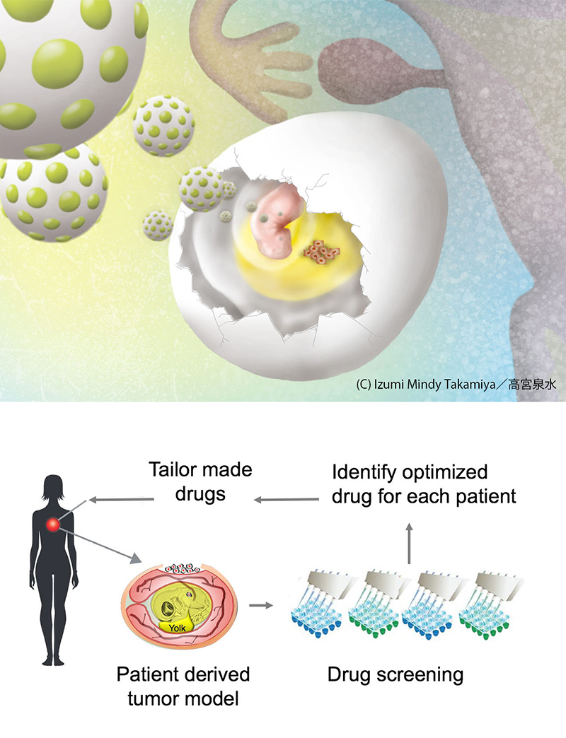
Successful replication of ovarian tumors inside chicken eggs heralds a new era for patient-centered cancer treatment.
Fuyuhiko Tamanoi of Kyoto University’s Institute for Integrated Cell-Material Sciences (iCeMS) and colleagues in the US succeeded in establishing a versatile, powerful and convenient model to analyze human cancer.
They developed a ‘chicken egg tumor model’ in which cultured ovarian cancer cells are transplanted on top of the membrane that surrounds a 10-day-old chicken embryo. An ovarian tumor forms on top of the membrane within three days of transplantation.
The team had similar results when they used ovarian tumor samples taken directly from patients, showing that their chicken egg model provides a convenient system for replicating human cancer. This conclusion is supported by their detailed characterization of the tumor, demonstrating that it possesses all major cancer features. “We were surprised when the tumor was formed in three days,” says Tamanoi. “This is very rapid considering that it takes weeks to do the same with mice. We can start using this model to test for anti-cancer drugs tailored to each cancer patient’s needs. The process can be completed within one week,” he says. This is a major step toward individualized medicine for cancer patients.
Tamanoi’s team, in collaboration with colleagues in France and Saudi Arabia, also developed a new type of biodegradable silica nanoparticle called ‘biodegradable PMO’, which is only 200 nanometers in size. The nanoparticles were loaded with the anti-cancer drug doxorubicin and were tested on human ovarian tumor established in the chicken egg.
The biodegradable PMO carrying doxorubicin quickly eliminated the human ovarian tumors without affecting other organs in the chicken embryo. When a smaller amount of the drug, not enveloped in the nanoparticles, was injected in the egg, severe organ damage ensued. This indicates that the team’s nanoparticles prevent anti-cancer drug side effects due to their ability to directly target the tumor.
The chicken egg model has several advantages over existing models, such as mouse models, for testing anti-cancer therapies. The tumors form much more rapidly on the chicken embryonic membranes than in mice due to the rich nutrient environment and the incomplete immune system at this stage of embryonic development. Fertilized chicken eggs are also less expensive to use than immune-compromised mice making the model suitable for high throughput experiments.
Paper information
【DOI】 https://doi.org/10.1038/s41598-018-25573-8
【KURENAI ACCESS URL】 http://hdl.handle.net/2433/231401
Binh Thanh Vu, Sophia Allaf Shahin, Jonas Croissant, Yevhen Fatieiev, Kotaro Matsumoto, Tan Le-Hoang Doan, Tammy Yik, Shirleen Simargi, Altagracia Conteras, Laura Ratliff, Chiara Mauriello Jimenez, Laurence Raehm, Niveen Khashab, Jean-Olivier Durand, Carlotta Glackin, Fuyuhiko Tamanoi (2018). Chick chorioallantoic membrane assay as an in vivo model to study the effect of nanoparticle-based anticancer drugs in ovarian cancer. Scientific Reports, 8, 8524.


