June 5, 2013

From left, Assoc. Prof. Kabashima, Postdoc fellow Egawa
Associate Professor Kenji Kabashima (Department dermatology, Graduate School of Medicine), Professor Yoshiki Miyachi, and Postdoctoral fellow Gyohei Egawa, all from Graduate School of Medicine, developed a new strategy to study vascular permeability in vivo.
This study was published in the "Scientific reports" on 4th June, 2013.
Outline
Blood vessel endothelium forms a semi-permeable barrier and its permeability controls the traffics of plasma contents. Here we report an intravital evaluation system for vascular permeability in mice using two-photon microscopy. We used various sizes of fluorescein-conjugated dextran as a tracer and its efflux was quantified by measuring the changes of fluorescent intensity both on the blood vessel area and the interstitial space. Using this system, we demonstrated that skin blood vessels limited the passage of dextran larger than 70 kDa under homeostatic conditions. We evaluated the kinetics of vascular permeability in histamine- or IgE-induced type I allergic models and a hapten-induced type IV allergic model. In such inflammatory conditions, the hyperpermeability was selectively induced in the postcapillary venules and dextran as large as 2000 kDa leaked from the bloods. Taken together, our study provides a convenient method to characterize the skin blood vessels as a traffic barrier in physiological conditions.
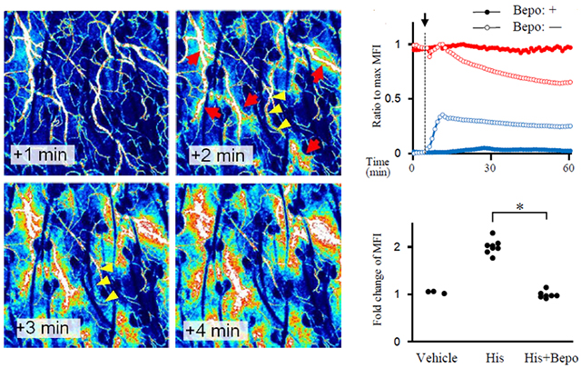
Figure:(Left)Sequential images after histamine injection. (Right)Kinetics of the MFI in the blood vessel area(red) and in the interstitial space(blue). The arrow denotes the timepoint of histamine injection. Mice were pretreated with bepotastine(closed circles) or vehicle(open circles).
Paper information
[DOI] http://dx.doi.org/10.1038/srep01932
Egawa Gyohei, Nakamizo Satoshi, Natsuaki Yohei, Doi Hiromi, Miyachi Yoshiki, Kabashima Kenji.
Intravital analysis of vascular permeability in mice using two-photon microscopy.
Scientific Reports 3, Article number: 1932, 2013/06/04/online